


Echocardiogram

What is Echocardiogram
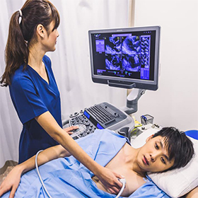
An Echocardiogram is a non-invasive diagnostic test that uses ultrasound waves to create moving images of the heart. It provides detailed information about the heart’s size, structure, and function, helping doctors evaluate heart conditions, detect abnormalities, and monitor treatment.
Key Features of an Echocardiogram:
- Ultrasound Technology: It uses high-frequency sound waves (ultrasound) to produce real-time images of the heart, which are displayed on a monitor.
- Heart Function Assessment: It allows the doctor to evaluate the heart’s pumping ability, how well the heart valves are working, and whether there are any areas of the heart muscle that aren’t functioning properly.
- Non-Invasive and Safe: The procedure involves no radiation and is generally safe, pain-free, and well-tolerated by patients.
- Variety of Uses:
- Detects conditions such as heart valve problems, congenital heart defects, heart failure, and pericardial effusion (fluid around the heart).
- Monitors the heart’s response after a heart attack or surgery.
- Helps assess the ejection fraction, which indicates how well the heart pumps blood.
- Types of Echocardiograms:
- Transthoracic Echocardiogram (TTE): The most common type, where a gel is applied to the chest, and a transducer is moved over the skin to capture heart images.
- Transesophageal Echocardiogram (TEE): Involves a special probe inserted into the esophagus to get clearer images, especially in certain heart conditions.
- Stress Echocardiogram: This combines an echocardiogram with a stress test (such as exercise or medication-induced) to assess the heart’s function under stress.